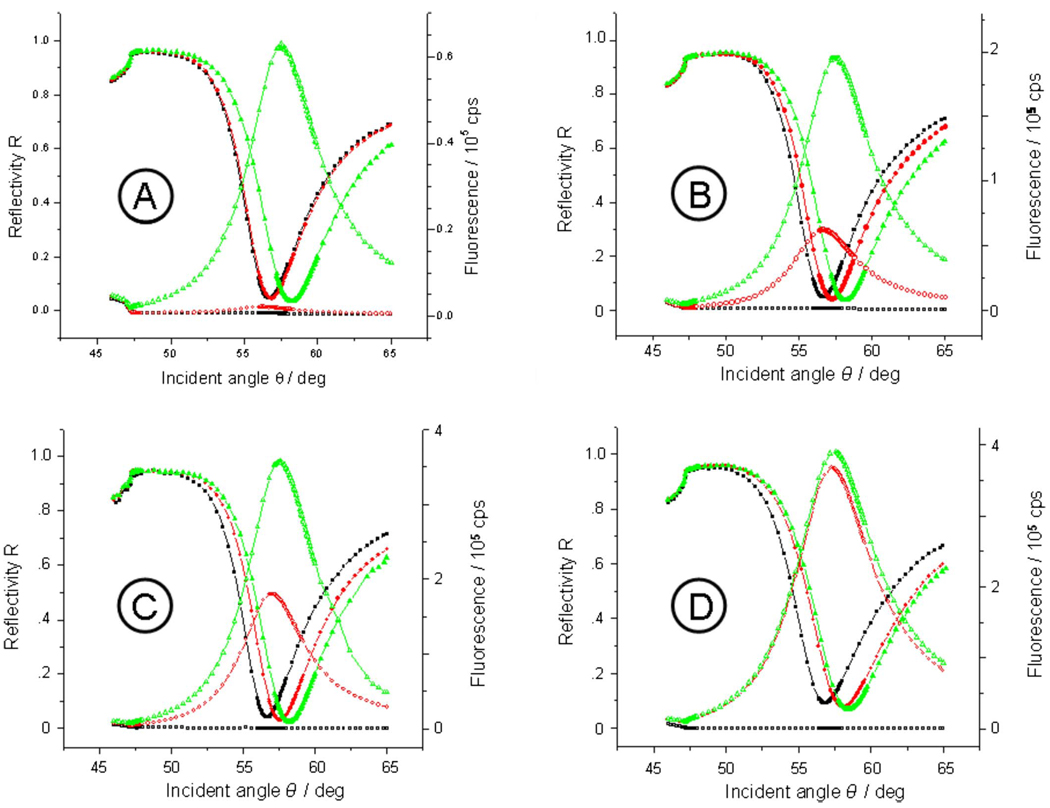
FIG. 2.
Angular dependences of the reflectivity (solid symbols) and the simultaneously measured fluorescence intensities (open symbols) for N-ELPs grafted on bare Cr/Au substrate with different graft densities. The black, red, and green curves represent the unmodified substrate prior to the injection of the ELP solution, after assembly of the labeled N-ELP brushes, and after the completion of the assembly process by unlabeled ELPs, respectively. The graft thickness of labeled N-ELP after step 1 is (a) d=1.7 nm, (b) 7.2 nm, (c) 11.0 nm, and (d) 12.9 nm, respectively.

