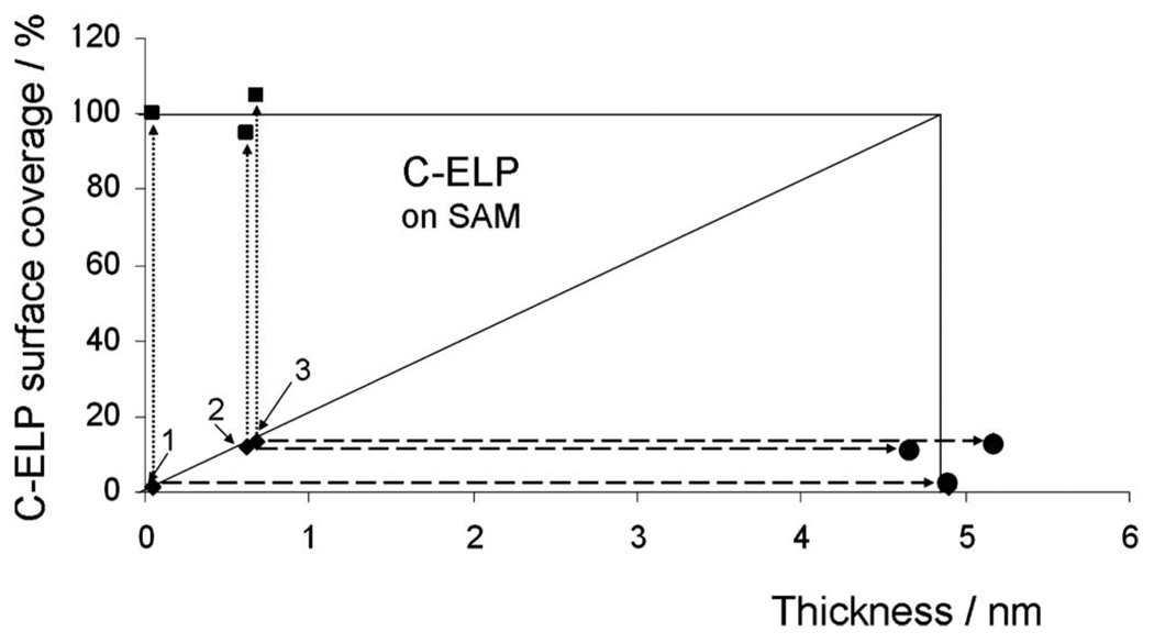
FIG. 6.
ELP surface coverage on COOH-(EG)6 NHS ester SAMs after the stepwise binding of labeled C-ELPs (♦) and unlabeled ELPs (■), respectively. The circle point (●) shows the final thickness after binding of unlabeled ELPs. The three initial C-ELP graft densities shown as 1, 2, and 3 are d=0.05 nm, 0.6 nm, and 0.7 nm, respectively.

