FIGURE 4.
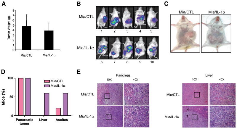
IL-1α secretion induces a metastatic phenotype in a pancreatic cancer orthotopic in vivo model. A. Tumors from mice injected with MiaPaCa-2/CTL or MiaPaCa-2/IL-1α cells were weighed. Columns, mean of all individual tumors in the group; bars, SE. B and C. Tumor development, as indicated by the Lumi-light, was monitored and photographed in real time as shown by the color. D. Percentage of mice that developed pancreatic tumors, liver metastasis, and ascites. E. H&E staining of the pancreas and liver tissues from SCID/NCr mice injected with MiaPaCa-2/CTL and MiaPaCa-2/IL-1α cells, either with ×10 or ×40 magnification. The square in ×10 magnification represents the field in the ×40 magnification. N, normal; T, tumor.

