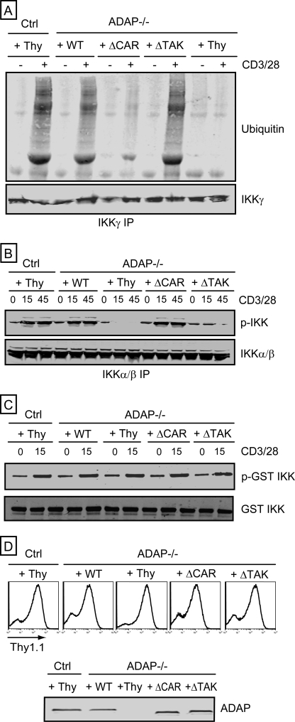
FIGURE 3.
Independent control of IKKγ ubiquitination and IKKα/β phosphorylation by ADAP. Control hCAR+ T cells (Ctrl) and hCAR+ ADAP−/− T cells were transduced with adenovirus encoding Thy1.1 alone (Thy) or wild-type ADAP (WT), the ADAP ΔCAR mutant, or the ADAP ΔTAK mutant prior to CD3/CD28 stimulation for 15 min (A and C) or for 15 or 45 min (B). A, IKKγ IPs were probed for ubiquitin and IKKγ. B, IKKα/β IPs were probed for phosphorylated IKK and IKK. C, in vitro kinase assays were performed with TAK1 IPs. Phosphorylation of GST·IKK was assessed by Western blotting with an anti-phospho-IKK antibody (p-GST IKK). Samples were also probed with an anti-IKK antibody (GST IKK). D, flow cytometry analysis of T cells infected with adenovirus with an anti-Thy1.1 antibody, which detects the Thy1.1 cell surface protein expressed by all recombinant adenoviruses used in this study. Cell lysates were also immunoblotted with an anti-ADAP antibody to confirm ADAP expression (bottom).

