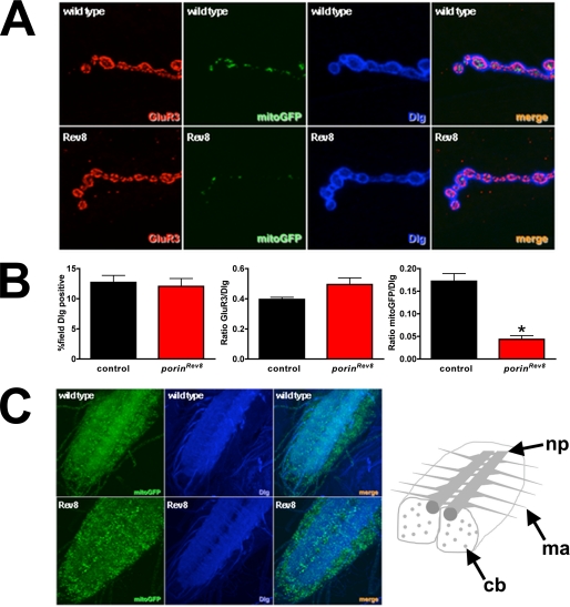
FIGURE 7.
porin mutants have reduced mitochondria in NMJ presynaptic termini. A, labeling of third instar larval NMJs with Dlg (blue) to outline boutons, GluR3 (GluRIII/IIC, red) to demarcate postsynaptic receptor field, and mitoGFP (green) expressed in motor neurons. B, quantification of fluorescence per field demonstrates decreased mitochondria in porinRev8. Error bars represent S.E. *, p < 0.05 by Student's t test with Welch correction. C, third instar larval ventral nerve cords labeled with Dlg (blue), which labels the neuropil (np) and motor axons (ma), and with mitoGFP (green). In porinRev8 animals, mitoGFP is more prominent in the cell bodies (cb) and sparser in the neuropil and motor axons compared with wild type (yw). Rev8, porinRev8.

