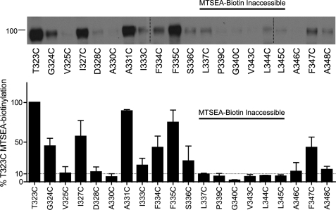
FIGURE 4.
Aqueous exposure of TM6 Cys mutants as revealed by MTSEA biotinylation. Accessibility of functional TM6 Cys mutants was assessed by application of 1 mm MTSEA-biotin and a subsequent Western blot using hSERT monoclonal antibody ST51–2. hSERT Cys mutants cDNAs were transiently transfected in HEK-293T cells at equal concentrations (see “Experimental Procedures”). Vertical dashed lines represent separate acrylamide gels run concurrently. Quantification of protein expression (n = 3) performed in Image J and normalized to T323C. The horizontal dashed line indicates 10% T323C MTSEA biotinylation.

