FIGURE 5.
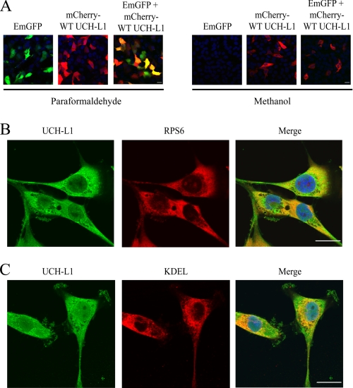
UCH-L1 is partially co-localized to the site of CFTR protein synthesis. A, UCH-L1 remains associated with cellular membranes following methanol fixation, whereas free cytosolic GFP leaks from the cells. IB3-1 cells were transiently transfected with EmGFP, mCherry-WT UCH-L1, or both. The cells were fixed in paraformaldehyde (cytosolic proteins retained) or methanol (cytosolic proteins leak). Blue indicates nuclear 4′,6′-diamino-2-phenylindole staining. The cells were analyzed using a Zeiss 510 Meta confocal microscope, and the images were processed using Zeiss LSM image examiner software. All of the images were obtained using identical confocal laser and scan settings. B, confocal microscopy demonstrates UCH-L1 partially co-localizes with the ribosome. IB3-1 cells were plated on 10-cm2 Mattek dishes. The cells were fixed in fresh 4% paraformaldehyde for 25 min, permeabilized in 0.2% Triton X-100 for 15 min, and blocked in 2% bovine serum albumin with PBS for 40 min. UCH-L1 and RPS6 (small ribosomal subunit) primary antibodies were added followed by Alexa 488 and 568 secondary antibodies. The nucleus was stained with 4′,6′-diamino-2-phenylindole. C, co-localization of UCH-L1 and the ER were examined by confocal microscopy as described in B. An antibody specific for the ER retention signal, KDEL, was used to immunostain the ER. The scale bar is equal to 20 μm.

