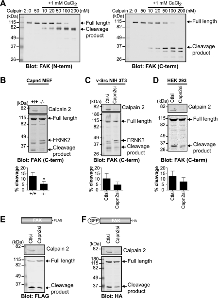
FIGURE 2.
FAK is cleaved by calpain 2 in vitro and in vivo. A, in vitro calpain cleavage assay of FAK was performed by incubating FAK in cleavage buffer alone or with calpain 2 or with 1 mm CaCl2 and increasing concentrations of calpain 2. Cleavage reactions were analyzed by immunoblotting and probed with anti-FAK antibodies specific to the N-terminal (N-term) and C-terminal (C-term) regions of FAK. Immunoblots shown are from one of two independent experiments. B–D, cell lysates from wild-type (+/+) and calpain 4 knock-out (−/−) mouse embryonic fibroblasts (Capn4 MEF) (B); v-Src-transformed NIH 3T3 control (Ctlsi) and calpain 2 (Capn2si) siRNA fibroblasts (C); and HEK 293 parental, control, and calpain 2 siRNA cells (D) were analyzed by immunoblotting and probed for calpain 2 and FAK. Immunoblots shown are from one of three independent experiments. Quantification of percent cleavage is defined as the ratio of cleaved FAK to total FAK (full-length + cleaved). Percent cleavage is shown as the mean ± S.E. *, p < 0.05 (by t test) compared with the control. E and F, cell lysates from HEK 293 control and calpain 2 siRNA cells transiently transfected with FAK-FLAG (E) or GFP-FAK (F) were analyzed by immunoblotting and probed with anti-calpain 2 and anti-FLAG (E) or anti-HA (F) antibodies. Immunoblots shown are from one of three independent experiments.

