FIGURE 5.
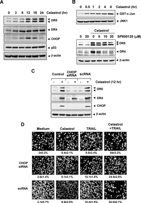
Up-regulation of DR5 by celastrol requires CHOP. A, MDA-MB-231 cells (5 × 10⁵) were incubated with 3 μmol/liter celastrol for the indicated times, and whole cell lysates were subjected to Western blotting analysis using relevant antibodies. B, upper panel, cells were incubated with 3 μmol/liter celastrol for indicated times, and whole cell extracts were immunoprecipitated with anti-JNK1 antibody and subjected to kinase assay as described under “Experimental Procedures.” The same protein extracts were subjected to Western blotting analysis using anti-JNK1 antibody. Lower panel, cells were pretreated with JNK inhibitor (SP600125) for 1 h and then exposed to 3 μmol/liter celastrol for 24 h. Whole cell extracts were prepared and analyzed for the expression of DR4 and DR5 using relevant antibodies. C, MDA-MB-231 cells (3 × 10⁶/well) were transfected with either CHOP siRNA or control siRNA. Twenty-four hours after the transfection, cells were re-seeded in 6-well plates or chamber slides. Cells were treated with 3 μmol/liter celastrol for 24 h, and whole cell lysates were analyzed by Western blotting (C). The same blots were stripped and reprobed with β-actin antibody to verify equal protein loading. D, cells were exposed to 2 μmol/liter celastrol for 6 h, washed with PBS to remove celastrol, and then treated with 10 ng/ml TRAIL. Cell death was determined by Live/Dead assay.

