FIGURE 1.
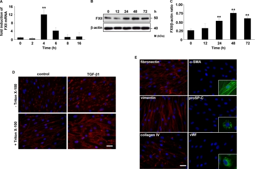
TGF-β1 up-regulates FXII expression in HLF. A and B, time course of FXII expression in HLF following TGF-β1 stimulation as assessed by real time PCR (A) and Western blotting (B). Real time PCR results are expressed as the fold increase in FXII expression (normalized for β-actin expression) versus values obtained for unstimulated cells and are means ± S.D.; n = 3; **, p < 0.01. The Western blot illustrated is from one representative experiment out of four. C, densitometric analysis of B. Data are presented as means ± S.D.; n = 4; **, p < 0.01, all versus unstimulated cells. D, immunofluorescence for the detection of FXII in unstimulated (control) or TGF-β1-treated HLF. Original magnification was ×40/1.25–0.75 oil objective. Bar size, 10 μm. E, immunofluorescence staining of HLF for fibronectin, vimentin, collagen IV, α-smooth muscle actin, pro-surfactant protein C, and von Willebrand factor. Original magnification was 40×/1.25–0.75 oil objective. Bar size, 10 μm. The insets are controls and show the positive staining of pulmonary artery smooth muscle cells for α-smooth muscle actin, of alveolar type II cells for pro-surfactant protein C, and of pulmonary artery endothelial cells for von Willebrand factor (vWf). α-SMA, α-smooth muscle actin; proSP-C, pro-surfactant protein C.

