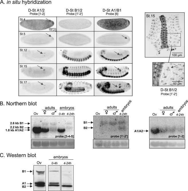
FIGURE 3.
Expression of the various D-stathmins during development. A, in situ hybridization with probes 1-2, 1′-2′, and 6 revealing various D-stathmin mRNA transcripts during embryonic development at different stages (St. 4, 5, 12, 15, and 17): D-stathmins A1/A2 are present in germ cells (arrows) and D-stathmin B1/B2 in the nervous system. Right panel: a higher magnification of the dissected nervous system at stage 15 labeled with probe 1′-2′ and showing the presence of D-stathmin B1/B2 in the sensory organs (arrow). B, Northern blot analysis of adult ovary (Ov) and adult and embryonic flies, with cDNA probes 3-4-5, 1′-2′, and 1-2, which detect all D-stathmin mRNAs, D-stathminS B1/B2, or D-stathmin A1/A2, respectively. The amount of RNA loaded on the gel was followed with methylene blue staining of rRNAs (bottom). C, D-stathmin Western blot analysis (antiserum 98) of adult ovary (Ov) and embryos.

