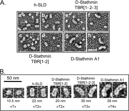
FIGURE 7.
Electron microscopy visualization of tubulin complexes with stathmin constructs. A, rotary metal shadowed electron micrographs of tubulin in the presence of the SLD of h-stathmin 4a (a and b), the TBR1-2 (c and d), and TBR1-2-3 (e and f) of D-stathmin constructs and D-stathmin A1 (g and h). In each field, uncomplexed individual tubulin molecules can be seen (arrowheads) as well as elongated complexes of increasing sizes (arrows). B, enlarged views of the various tubulin complexes shown in A, with their calculated “uncoated” lengths (see “Experimental Procedures”) and deduced tubulin stoichiometries, clearly showing the formation of T4S complexes with D-stathmin A1.

