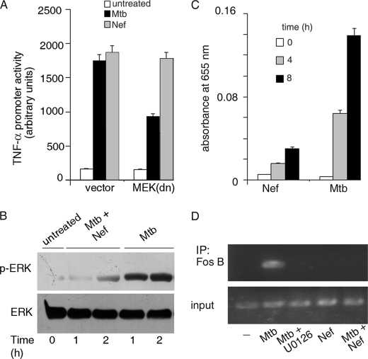
FIGURE 4.
Role of MAPKs in the activation of TNF-α. A, THP-1 cells were transfected with empty vector or dominant-negative MEK (MEK(dn)) along with the TNF-α promoter luciferase reporter construct. Transfected cells were left untreated or treated with Mtb or with Nef, and luciferase activity was measured as described previously. B, THP-1 cells were left untreated or treated with Mtb in the absence or presence of Nef for different periods of time. Cell lysates were immunoblotted with phospho-ERK antibody, and blots were reprobed with ERK antibody. C, THP-1 cells were left untreated or treated with Mtb or Nef for the indicated periods of time (in hours), and the activation of FosB was quantified using the TransFactor ELISA kit (Clontech) according to the manufacturer's protocol. D, THP-1 cells were left untreated (−) or preincubated with the inhibitor U0126 prior to treatment with Mtb. In a separate set of experiments, cells were treated with either Mtb or Nef or with both. After treatments, ChIP analysis was carried out with primers specific for the AP1-binding site of the TNF-α promoter after immunoprecipitation (IP) with anti-FosB antibody. The input panel shows the PCR product obtained when no immunoprecipitation was performed.

