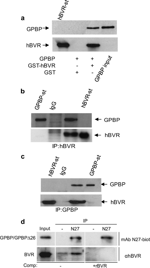
FIGURE 1.
GPBP binds to hBVR in HEK293A cells. a, a GST pulldown assay is shown. HEK293A cells were transfected with the pcDNA3-expression plasmid encoding GPBP. One day after DNA addition, cell lysates were prepared and subjected to a GST pulldown assay using either GST-hBVR or GST alone. The membrane was sequentially probed with anti-GPBP and anti-hBVR antibodies. b, co-IP of GPBP and hBVR detected by hBVR antibody is shown. Lysate prepared from cells co-transfected with pcDNA3-hBVR and pcDNA3-GPBP was immunoprecipitated with an anti-hBVR antibody or with control rabbit IgG. The precipitate was subjected to Western blot analysis by sequentially probing the membrane with anti-GPBP and anti-hBVR antibodies. c, co-IP of GPBP and hBVR as detected by GPBP antibody is shown. The same cell lysates were immunoprecipitated using anti-GPBP antibody or control rabbit IgG and analyzed as in b. d, cross-linking of GPBP and hBVR is shown. HEK293A cells were cross-linked with formaldehyde, and cell extracts were immunoprecipitated with protein A-Sepharose after incubation with or without the indicated GPBP-specific antibodies. The input lane shows the presence of both GPBP (and/or the closely migrating GPBPΔ26) and BVR. The immunoprecipitates were resolved by gel electrophoresis, and the Western blot was probed with biotinylated N27 antibody or anti-BVR. In the competition experiment, anti-BVR binding was blocked by the addition of recombinant hBVR. In a–d, detection was by secondary antibody conjugate followed by ECL. Purified preparations of GST-hBVR (BVR-st) and GPBP were included as controls (GPBP-st).

