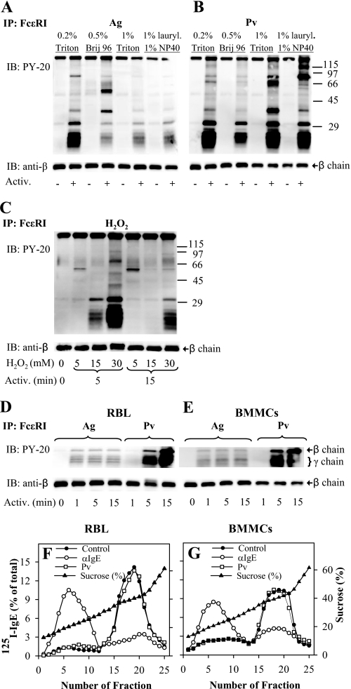
FIGURE 1.
Tyrosine phosphorylation of FcϵRI in the absence of its association with DRMs. A, IgE-sensitized RBL cells were activated (Activ.; +) or not (−) for 5 min with 0.5 μg/ml TNP-BSA (Ag). After activation, the cells were solubilized in lysis buffers containing 0.2% Triton X-100 (Triton), 0.5% Brij 96, 1% Triton X-100, or 1% n-dodecyl β-d-maltoside (lauryl.) and 1% Nonidet P-40 (NP-40). FcϵRI receptor was immunoprecipitated (IP) from postnuclear supernatant using anti-IgE-armed protein A beads. The immunocomplexes were size-fractionated by SDS-PAGE and analyzed by immunoblotting (IB) with phosphotyrosine-specific antibody-HRP conjugates (PY-20). After stripping, the membranes were probed with antibody specific for FcϵRI-β subunit (anti-β), which served as a loading control. B, IgE-sensitized RBL cells were activated (+) or not (−) for 5 min with 0.2 mm Pervanadate (Pv), solubilized, and analyzed as in A. Numbers on the right indicate positions of molecular mass markers (kDa). C, IgE-sensitized RBL cells were either not activated (0 min) or activated for 5 or 15 min with different concentrations (5–30 mm) of H2O2, then solubilized in lysis buffer containing 0.2% Triton X-100, and analyzed as in A. D, IgE-sensitized RBL cells were activated for different time intervals with 0.5 μg/ml TNP-BSA (Ag) or 0.2 mm Pv, then solubilized in lysis buffer containing 0.5% Triton X-100, and analyzed as in A. E, IgE-sensitized BMMCs were treated and analyzed as in D; only FcϵRI β and γ subunits are shown (on the right). F and G, sucrose gradient ultracentrifugation of cell lysates. 125I-IgE-sensitized RBL cells (F) and BMMCs (G) were exposed to anti-IgE (αIgE), 0.2 mm Pv, or BSS/BSA alone (Control). After 5 min, the cells were solubilized in lysis buffer containing 0.06% Triton X-100. Lysates were then diluted 1:1 with 80% sucrose buffer, loaded into sucrose step gradients, and ultracentrifuged. After fractionation, 0.2-ml aliquots were collected from the top of the gradient, and the distribution of 125I-IgE-FcϵRI complexes was expressed as percentage of total radioactivity present in individual fractions. Percentage of sucrose in the fractions was determined with Abbe refractometer. Representative data from two (C) or three (all other) experiments are shown.

