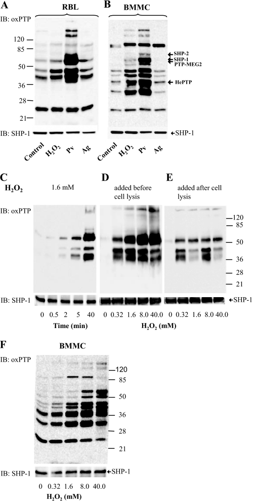
FIGURE 3.
Numerous PTPs are oxidized in the course of mast cell activation. A and B, IgE-sensitized RBL cells (A) or BMMCs (B) were nonactivated (Control) or activated with 5 mm H2O2, 0.2 mm Pv, or TNP/BSA (Ag; 0.5 μg/ml). After 15 min, the cells were solubilized in lysis buffer containing 0.2% Triton X-100. Postnuclear supernatants were size-fractionated by SDS-PAGE and analyzed by immunoblotting (IB) with mAb specific for the oxidized PTP active site. After stripping, the membranes were probed with anti-SHP-1 antibody as a loading control (SHP-1). Numbers on the left and right indicate, respectively, position of molecular mass markers (in kDa) and localization of phosphatases as identified by immunoprecipitation and/or mass spectrometry (see supplemental Fig. S2 and Tables S1 and S2). C, RBL cells were activated with 1.6 mm H2O2 for the indicated time intervals. The cells were lysed and processed as in A. D, RBL cells were activated for 40 min with different concentrations of H2O2. Subsequently, the cells were lysed in 0.2% Triton X-100 and processed as in A. E, RBL cells were first lysed with 0.2% Triton X-100, and the postnuclear supernatants were exposed to H2O2 at the indicated concentrations. After 40 min, the lysates were fractionated by SDS-PAGE and analyzed as in A. F, BMMCs were activated for 40 min with different concentrations of H2O2. Subsequently, the cells were lysed in 0.2% Triton X-100 and processed as in A. Typical experiments from at least three performed are shown.

