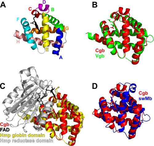
FIGURE 1.
Backbone topology of Cgb. A, Cα chain tracing of Cgb with heme cofactor (black). Helices/regions are labeled according to conventional globin nomenclature. B–D, overlays of the Cgb backbone (red) with Vgb from Vitreoscilla stercoraria (Protein Data Bank entry 1VHB (14)) (B), N-terminal domain of Hmp from E. coli (Protein Data Bank entry 1GVH (6)) (C), and sperm whale myoglobin (Protein Data Bank entry 2JHO (36)) (D).

