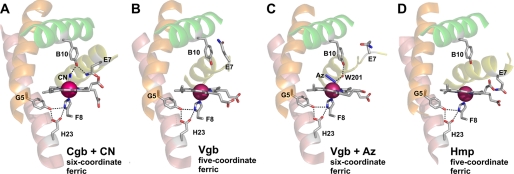
FIGURE 4.
The active site structure of Cgb (A), Vgb (Protein Data Bank entry 1VHB) (B), azide-bound Vgb (Protein Data Bank entry 2VHB) (C), and Hmp (Protein Data Bank entry 1GVH) (D). The B, E, G, and H helices are colored green, yellow, orange, and salmon, respectively. Bound cyanide and azide are labeled CN and Az, respectively, and hydrogen bonding interactions inferred from the structures are indicated by dotted lines.

