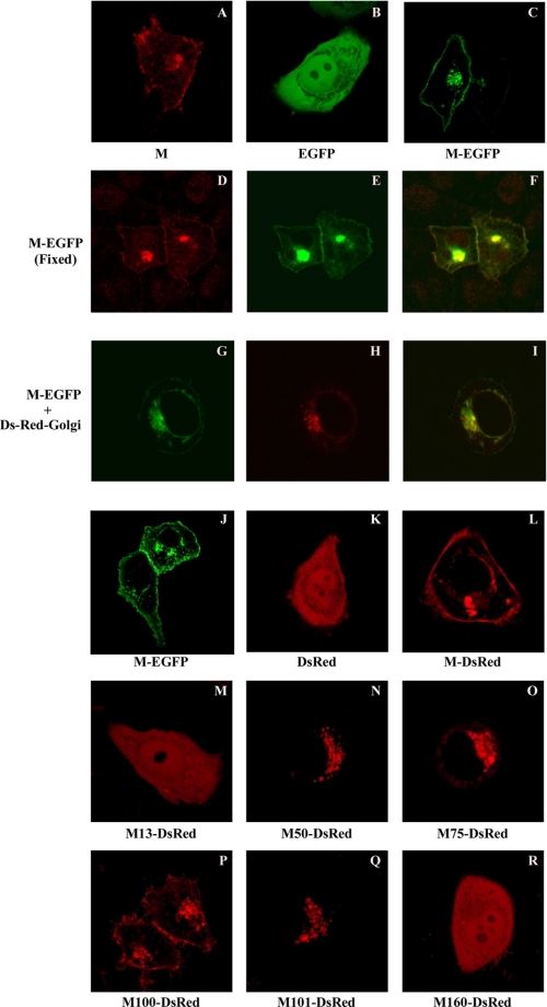
FIGURE 6.
Subcellular localization of SARS-CoV M (untagged or tagged with a fluorescent protein) in fixed or living cells. HeLa (A–F and K–R), 293T (G–I), or Vero-E6 (J) cells were transfected or cotransfected with the indicated expression vectors. pM-EGFP and pM-DsRed encode SARS-CoV M bearing carboxyl-terminal-tagged EGFP and DsRed, respectively. pDs-Red-Golgi encodes a Golgi apparatus labeling marker. At 4 h (G–I) or 24 h post-transfection, cells were either fixed or directly observed using a laser confocal microscope. Fixed cells (A and D–F) were labeled with a primary anti-SARS-CoV M antibody and a secondary rhodamine-conjugated anti-rabbit antibody. Images shown here represent the most prevalent phenotypes. Merged red and green fluorescence images (D and E) are shown in F. Superimposed fluorescence and phase-contrast images (G and H) are shown in I. Mock-transfected cells failed to yield any signal (data not shown).

