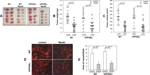
FIGURE 5.
Ligand activation of human PPARα in PPARα humanized (hPPARα) mice blunts tumor angiogenesis and growth. Groups of WT and hPPARα mice were either left untreated or administered Wyeth (0.02%, v/v) in their drinking water for 2 days prior to receiving two subcutaneous injections with p60.5 cells. Wyeth treatment was continued for the next 2 weeks, at which point mice were sacrificed, and their tumor load was quantified. A, representative images of tumors grown in untreated and Wyeth-treated WT and hPPARα mice. B and C, quantification of the weight (B) and volume (C) of tumors grown in untreated and Wyeth-treated WT and hPPARα mice. Circles show individual tumor values, whereas bars show mean values. D and E, frozen sections of tumors from untreated (control) and Wyeth-treated (Wyeth) WT and hPPARα mice were stained with anti-mouse CD31 antibodies (D), and their degrees of vascularization was quantified as the percentage of the area occupied by CD31-positive structures per microscopic field (E). The values in panel E are averages ± S.D. calculated from ten tumors/group with two images analyzed per tumor.

