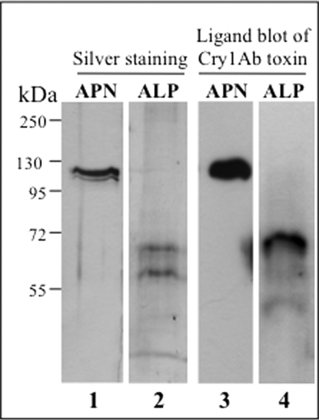
FIGURE 2.
Purification of M. sexta APN and ALP proteins. Silver stain 10% SDS-PAGE of APN (lane 1) and ALP (lane 2) pure fractions from Mono Q (Table 1) and affinity chromatography (Table 2), respectively, are shown. Ligand blots with biotin-labeled Cry1Ab toxin of APN (lane 3) and ALP (lane 4) fractions are shown. Molecular mass markers are shown at the left of the figure.

