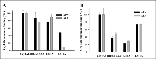
FIGURE 5.
Binding analysis of Cry1Ab and loop 2 and domain III mutants to APN or ALP. A, ELISA binding assays of 25 nm of monomeric structures of Cry1Ab and mutant toxins to APN (black bars) or ALP (gray bars). B, ELISA binding assays of 0.1 nm of oligomeric structures of Cry1Ab and mutant toxins to APN (black bars) or ALP (gray bars). Standard deviations from three replica plates were obtained.

