
Figure 2.
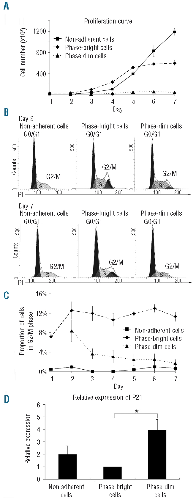
Cell proliferation and cell cycle status. (A) Proliferation kinetics of the three cell fractions (N=4). (B) Representative propidium iodide (PI) staining of cells from the three distinct compartments at day 3 and 7. (C) Dynamics of the three cell fractions in G2/M phase during 1 week of co-culture (N=3). (D) Relative expression of p21 in the three compartments at day 5 (N=3, *P<0.05). The data were normalized to the p21 expression in phase-bright cells, which was arbitrarily set at one.