
Figure 1.
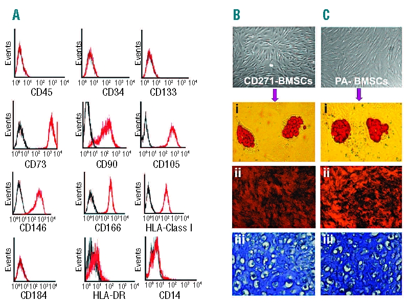
Phenotype of ex vivo expanded CD271- MSC and their in vitro differentiation potential. (A) CD271-MSC express high levels of all typical MSC markers such as CD73, CD90, CD146, CD166 and CD105. They also express HLA- class I antigens but do not express HLA-DR, CD45, CD14, CD184, or hematopoietic stem cells markers (CD34 and CD133). (B) Differentiation potential of CD271-MSC and PA-MSC (C). Like PA-MSC (Ci-iii) these MSC differentiate into adipocytes (Bi), osteoblasts (Bii) and chondrocytes (Biii). Accumulation of intracellular lipid vacuoles was shown by oil red-O staining for 18 days in culture with NH AdipoDiff medium, while osteoblast differentiation was detected by alkaline phosphatase activity after 10 days in NH OsteoDiff medium. Cartilage matrix deposition along with chondrocytes in lacunae was demonstrated by metachromatic toluidine blue staining after 24 days in NH ChondroDiff medium. Magnification for microphotographs of MSC and osteoblasts was 20x; the magnification for adipocytes was 200x and for chondrocytes 400x.