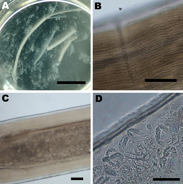
Figure.
Images of the adult female Dirofilaria repens worm removed from a subcutanous nodule of the patient. A) Macroscopic view of sections of the worm in saline in a petri dish. Two uteri and the intestinal tract can be seen protruding from a disrupted end of the largest section. The saline is turbid due to the massive release of microfilariae, which are not discernible at this magnification. Scale bar = 1 cm. B) Microscopic view of the outer cuticula with multiple longitudinal ridges. Scale bar = 100 μm. C) Microscopic view of the worm showing the well-developed muscle layer and the uterus containing microfilariae. Scale bar = 100 μm. D) Higher magnification of a section of the uterus containing multiple microfilariae. Scale bar = 50 µm.

