
Figure 3. Inhibition of caspase dependent cell death in prostatic acini.
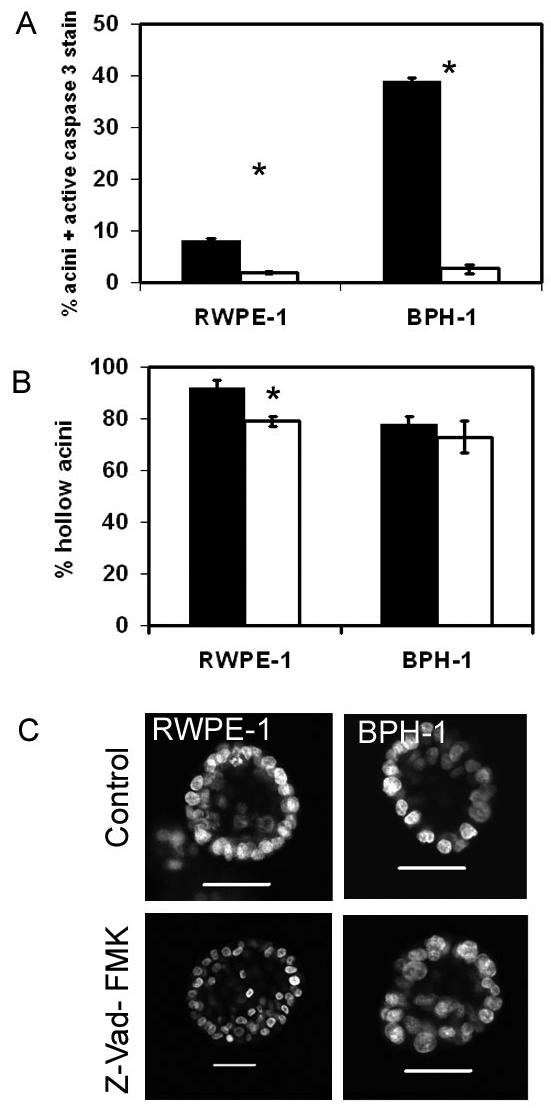
Prostate cells were grown in Matrigel for 2 days and then grown in the presence of 100 μM Z-VAD-FMK (open) or DMSO control (filled). Acini were grown for a further 4 days and then fixed and stained for active caspase 3, nuclei were counterstained with DAPI. A hundred acini were counted for the expression of active caspase 3 (A) or the presence of lumen (B). The graphs indicate the results of 3 replicate experiments ± sem, * p<0.015 (students t-test). Representative images of RWPE-1 or BPH-1 cells grown with or without caspase inhibitor, and nuclei were counterstained with DAPI are shown in (C). All bars indicate 50 μm. Images are cross-sections through the middle of acini.