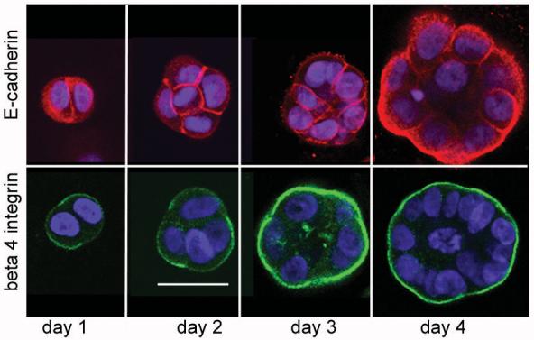
Figure 5. Polarisation of E-cadherin and beta 4 integrin in BPH-1 epithelial acini.

BPH-1 cells were grown in Matrigel for the indicated number of days. Acini were then fixed and stained for E-cadherin (red) or beta 4 integrin (green), nuclei were counterstained with DAPI. Representative images (of 3 independent experiments) are shown that cross section through the middle of cells or developing acini. Images were taken at x20 magnification, the bar indicates 50 μm.
