
Figure 6. Ouabain and anti-Na+/K+ATPase inhibition of lumen formation in prostatic acini.
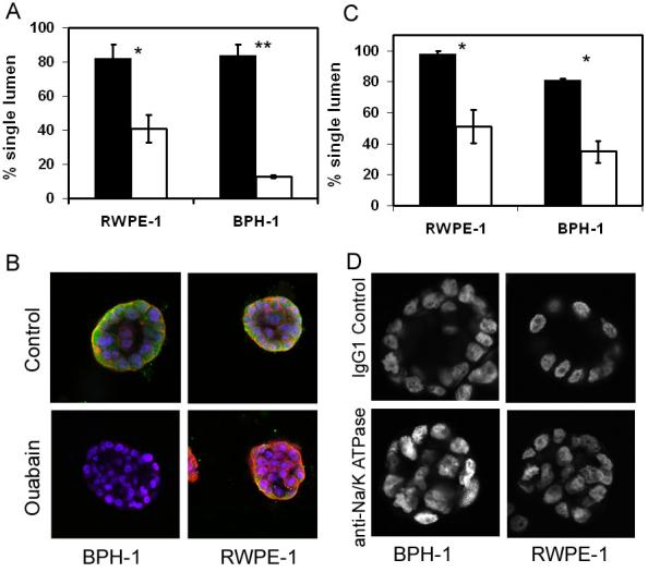
A. RWPE-1 or BPH-1 cells were grown in 3D Matrigel culture for 4 days and then treated with 1 nM ouabain (open) or DMSO control (filled). After 24 hours culture 50 acini were counted for the presence of single lumen. B. Acini were stained for E-cadherin (red) and PSA (green), nuclei were counterstained with DAPI. Images were taken at x20 magnification.
C. Cells were plated into 3D Matrigel and treated with 1/100 IgG1 control (filled) or 1/100 anti-Na+/K+ ATPase (open) for 6 days. 50 acini were counted for the presence of single lumen. All graphs indicate the results of 3 replicate experiments ± sem, * p<0.05, **p<0.0003 (students t-test). D. Acini were stained with DAPI and mid-section images were taken at x20 magnification.