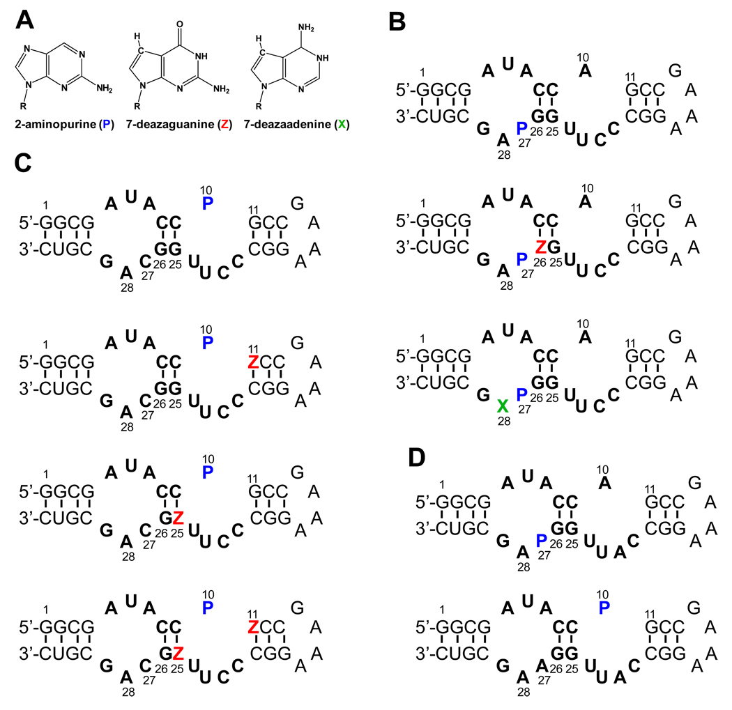
Figure 2.
(A) Structures of 2-aminopurine (P), 7-deazaguanine (Z), and 7-deazaadenine (X). Sequences and secondary structures of (B) the theophylline-binding aptamer RNA constructs with 2AP (P) labeled at residue 27; (C) the theophylline-binding aptamer RNA constructs with 2AP labeled at residue 10; and (D) 3MX-binding aptamer RNA constructs with 2AP at residues 27 and 10.

