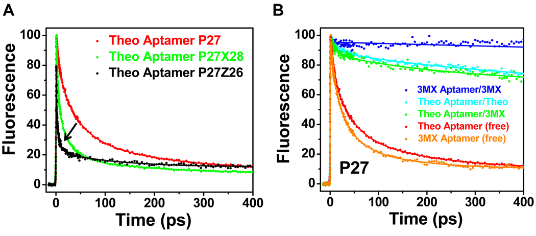
Figure 3.
Ultrafast decay profiles measured at the magic angle for the P27 constructs. (A) Decay profiles for the free theophylline-binding aptamer constructs P27 (red), P27Z26 (black), and P27X28 (green). The arrow indicates the change of dynamic timescale from slower in P27 to faster in P27Z26 and P27ZX28 (see text). (B) Comparison of decay profiles between free aptamer P27 constructs (theophylline, red; 3MX, orange) and complexes with theophylline (cyan) or 3MX (green and blue).

