
Figure 3.
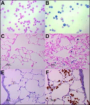
Immunohistochemistry for iron and ferritin in rat lung sections following NE or control vehicle intratracheal instillation. Sprague‐Dawley rats were exposed to NE (50 μg; B, D, F) or to control vehicles (A, C, E) via oral aspiration. Twenty‐eight days following exposure, rats were sacrificed and were lavaged (A, B) or lungs were harvested and fixed for histology and immunohistochemistry (C–F). BAL cell pellets (A, B) were stained with Perl's Prussian blue stain for iron. Lung sections were stained for iron (C, D) and for ferritin (E, F). Micrographs are representative of n= 4 animals. NE increased both iron staining and ferritin staining.