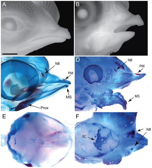
Figure 3. Embryos infected with RCAS-Bmp at HH11 produce large amounts of cartilage at the expense of bone.
(A) Side view of a normal embryo at day 13. (B) Embryos infected with RCAS-Bmp-2 or RCAS-Bmp-4 exhibit severe malformations. In this embryo, the lower jaw was truncated and dysmorphic. (C) Whole skeletal preparations stained with alcian blue (cartilage) and alizarin red (bone) reveal that normally at this time the nasal bone (NB), the premaxillary bone (PM), and bones in the proximal jaw (Prox) and the mandibular symphsis (MS) are evident. The cartilage elements of the upper and lower jaw are also in place. (D) In embryos infected with RCAS-Bmp-2 or Bmp-4 a large amorphous cartilaginous mass is apparent in the upper and lower jaw. The nasal bone (NB), premaxillary bone (pM) and the mandibular symphysis are rudimentary. (E) Dorsal view of the roof of the skull demonstrating that normally, the bones have not formed by this time. (F) Embryos infected with RCAS-Bmp-2 or Bmp-4 have ectopic cartilage (EC) covering the brain Scale bar: 250μm.

