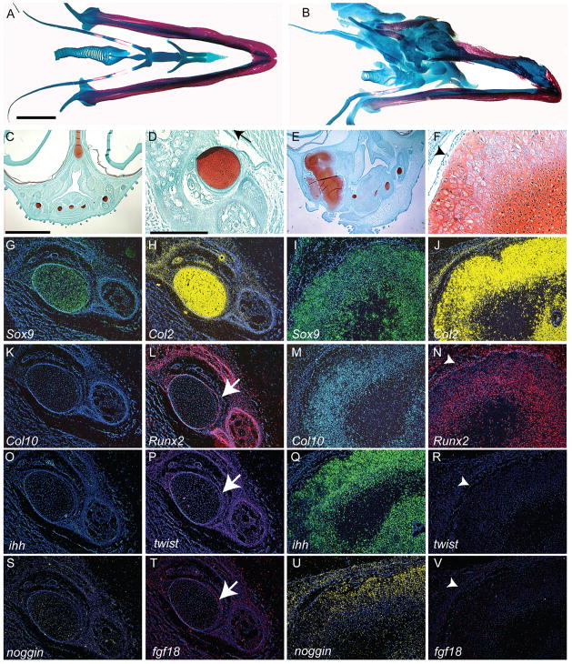
Figure 6. Morphological and molecular alterations of the jaw.
(A) Alcian Blue and Alizarin Red staining illustrates the cartilage (blue) and bone (red) elements comprising the tongue and mandible of chick embryos incubated for 15 days (n=3). (B) In treated embryos (HH22) incubated for 14 days (n=10), the jaw and tongue skeleton exhibit increased cartilage formation. (C) A transverse section, stained with safranin-o and fast green, through the tongue and lower jaw of an embryo incubated for 14 days illustrates the normal morphology of the cartilages (red) that comprise the skeleton. (D) Higher magnification of Meckel’s cartilage (red) of the lower jaw. (E) A section through a treated embryo reveals the large amount of cartilage that formed in response to BMP signaling. (F) Higher magnification of cartilage in E shows chondrocytes that appear to be hypertrophic. These cartilages do not have a well-formed perichondrium (arrowhead). (G) Sox9 (green) expression was normally restricted to the developing cartilages, where (H) Col2 (yellow) was also expressed. Col2 transcripts were also detected in mesenchymal cells adjacent to the cartilage. (I) In treated embryos the Sox9 and (J) Col2 expression domains were expanded, but their spatial patterns are not altered. (K) Normally, Col10 was not expressed by Meckel’s cartilage, and (L) Runx2 was only expressed in the adjacent bones and the perichondrium (arrow). (M) However, in the hypertrophic chondrocytes located in the ectopic cartilages, Col10 and (N) Runx2 transcripts were present. In the perichondrium (arrowhead), Runx2 transcripts were down-regulated in some regions, and not detected in others (not shown). (O) Ihh was not normally expressed in Meckel’s cartilage. (P) Twist-1 (purple) transcripts were detected in the perichondrium of normal Meckel’s cartilage. (Q) Ihh transcripts were detected in chondrocytes of treated cartilage. (R) Twist-1 was not detected in the perichondrium of treated embryos. (S) Normally, Noggin was not expressed by mandibular chondrocytes. (T) Fgf18 transcripts (pink) were detected in the perichondrium of Meckel’s cartilage. (U) After infection with RCAS-Bmp-4 Noggin expression was upregulated in chondrocytes. (V) Fgf18 transcripts were absent from the perichondrial region of treated cartilage. A,B,C,E: 250μm, D,F,G-V: 200μm

