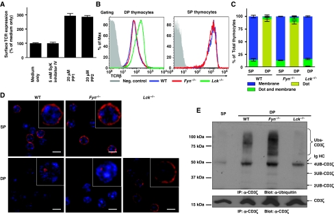
Figure 2.
Lck is required for tonic ubiquitylation of CD3 in DP thymocytes. (A) Thymic lobes from newborn mice (P1) were treated with the inhibitors indicated for 20 h, and surface TCRβ expression determined by flow cytometry. Data represent the mean±s.e.m. of 5–10 mice from 2–3 experiments per group. (B) Thymi were isolated from WT, Fyn−/−, or Lck−/− mice. Surface expression of the TCR:CD3 complex was measured by flow cytometry on thymocytes co-stained with mAbs against CD4, CD8, and TCRβ. (C, D) Thymocytes were stained with mAbs against CD4 and CD8, and DP and SP thymocytes purified by FACS. Sorted thymocytes were fixed, permeabilized, and stained with Alexa-647-conjugated CD3ζ mAb. The distribution of CD3ζ in thymocyte populations is indicated (n>50) (C). The localization of CD3ζ (red) and DAPI stained nucleus (blue) are shown in representative confocal images (D). (E) Tonic ubiquitylation of CD3 was performed with WT, Fyn−/−, or Lck−/− mice as described in Figure 1A.

