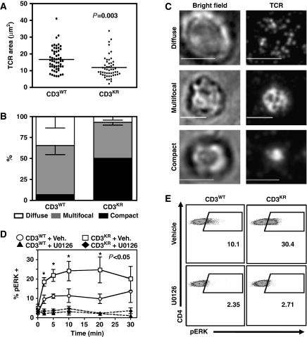
Figure 6.
Impaired CD3 ubiquitylation enhances IS formation and increases ERK phosphorylation in DP thymocytes. (A–C) DP thymocytes from CD3WT or CD3KR Rg mice were purified by FACS and added to a synthetic planar lipid bilayer containing unlabelled His-tagged ICAM-1 and fluorescently labelled streptavidin conjugated to biotinylated anti-TCRβ antibodies. Interactions were fixed following 15 min stimulation, visualized using spinning disk confocal microscopy and analysed using Slidebook software. (A) The area of individual DP T-cell synapses obtained in two independent experiments. (B, C) The morphology of individual synapses were classified according to the representative images (B) and shown graphically (C—Scale bar=5 mm). (D, E) Thymocytes from CD3WT or CD3KR Rg mice were isolated, rested in 0.5% FCS RPMI for 1 h at 37°C, and then activated by crosslinking using a CD3ɛ mAb in the presence (+U0126) or absence (+Veh.) of the MEK inhibitor U0126. Thymocytes were then fixed at the indicated time, permeablized, and stained with CD4, CD8, and pERK Abs. (D) CD4+CD8+ DP thymocytes were gated and pERK expression measured. (E) Representative flow cytometry dot plots are shown 10 min after stimulation. Statistical significance was determined using an unpaired t test in Prism software.

