Figure 7.
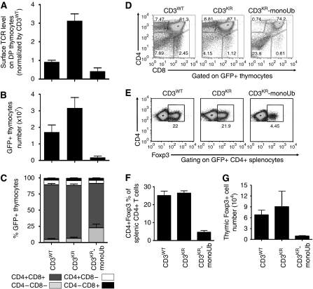
CD3 ubiquitylation alters TCR expression, thymic cellularity, and Treg development. Retrogenic mice were generated by reconstituting sublethally irradiated Rag1−/− recipients with transduced CD3ɛζ−/− bone marrow. Mice were analysed 5–8 weeks after transfer. Thymocytes were counted and stained with antibodies to CD4, CD8, and TCRβ, and analysed by flow cytometry. Surface TCR level on GFP+ DP thymocytes (A), GFP+ thymocyte number (B), and the percentage of GFP+ thymocytes (C) was determined. Data were gated on live, GFP+ cells, and represent the mean±s.e.m. of 10–20 mice from more than three experiments per group. (D) Representative dot plots of GFP+ thymocytes stained with antibodies to CD4 and CD8 are shown (representative of 10–20 mice from more than three experiments). (E–G) Splenocytes and thymocytes were surface stained with CD4 mAb and intracellularly stained with Foxp3 mAb. Representative dot plots of CD4+ splenic T cells from a retrogenic mouse experiment are shown (E). Bar charts show the percentage of splenic CD4+ T-cells expressing Foxp3 (F) and the number of Foxp3+ thymocytes (G). A full-colour version of this figure is available at The EMBO Journal Online.

