Figure 3.
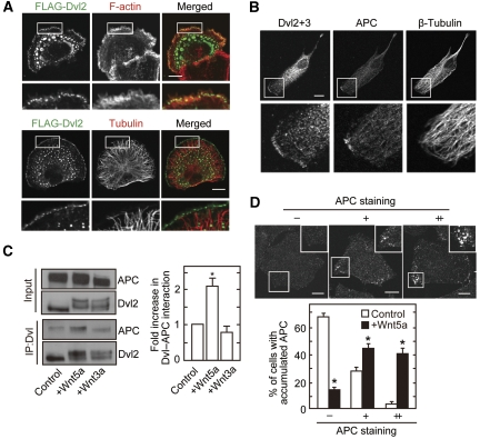
APC localizes to the cell periphery with Dvl in response to Wnt5a. (A) Vero cells expressing FLAG-Dvl2 were plated onto collagen-coated dishes for 1 h, followed by the staining with anti-FLAG antibody and phalloidin or anti-tubulin antibody. Cortical localization of FLAG-Dvl2 was observed in 42% of 30 cells expressing FLAG-Dvl2. (B) Vero cells were fixed with methanol and stained with anti-β-tubulin, anti-Dvl2+3 (a mixture of anti-Dvl2 and anti-Dvl3 antibodies), and anti-APC antibodies. Polarized Dvl at the cell cortex was observed in 42% of 96 cells and colocalization of Dvl with APC was observed in 27% of 41 cells with cortical Dvl staining. (C) After NIH3T3 cells were treated with Wnt5a or Wnt3a for 30 min, lysates were immunoprecipitated with anti-Dvl (DIX) antibody. The immunoprecipitates were probed with anti-APC and anti-Dvl2 antibodies, and band intensities of precipitated APC were quantified in the right-hand panel. (D) After HeLaS3 cells were serum-starved for 36 h and stimulated with or without 400 ng/ml purified Wnt5a for 60 min, the cells were stained with anti-APC antibody. Cells with a high intensity of APC at the cell periphery that was 1.5–2 times stronger than at the cell centre were defined as (+), and 2 times or more stronger were defined as (++). Cells with accumulated APC (−, +, ++) at the cell periphery was counted in at least 50 cells per each treatment. Scale bars in (A), (B), and (D), 10 μm. *P<0.01.

