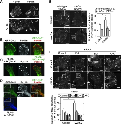
Figure 6.
The Dvl and APC complex stimulates the focal adhesion dynamics in response to Wnt5a. (A) Vero cells were stained with anti-paxillin antibody and phalloidin. The region in white boxes (a1 and a2) are shown enlarged in bottom panels. (B, C) Vero cells expressing GFP-Dvl2 (B) or FLAG-APC(Arm+) (C) were stained with the indicated antibodies. (D) Vero cells coexpressing FLAG-APC(Arm+) with GFP-Dvl2 were stained with anti-paxillin, anti-FLAG, and anti-GFP antibodies. The region in the white box is shown enlarged. (E, F) HeLaS3 cells expressing HA-Dvl1(DEP+) (E) or transfected with siRNA for Fz2, Dvl, or APC (F) were stimulated with Wnt5a conditioned medium for 60 min and then the cells were stained with anti-paxillin antibody. The region in the white box is shown enlarged. The numbers of focal adhesions in three different areas (15 μm × 15 μm) at the cell periphery (white box) were counted in 20 cells per treatment. Scale bars, 10 μm. *P<0.01.

