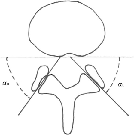
Fig. 2.
Each facet joint angle was measured using an axial T1-weighted image, which was scanned parallel to the lower end plate at the L4-L5 intervertebral disc space. The right and left facet joint orientations in the coronal plane were then measured as the angle between the line drawn tangentially to the posterior wall of the vertebral body and the line drawn through the interarticular gap at the medial and lateral extremities of the facet joint.

