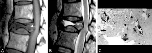
Fig. 1.
A 65-year-old woman with delayed post-traumatic vertebral collapse of T12 (pattern A, fluid pattern). (A) Spin echo T1-weighted (567/18) sagittal MR image shows a predominant area of hypo-intense signal intensity (arrow). (B) Fast spin echo T2-weighted (4,000/108) sagittal image obtained at the same level as in A shows a predominant area of hyper-intense signal intensity similar to water (arrow). (C) Corresponding photomicrograph of the specimen reveals extensive bone necrosis (arrows).

