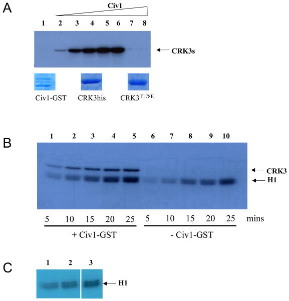
Figure 2. Phosphorylation of CRK3 with a CDK-activating kinase.
(A) Upper panel. Phosphorylation of CRK3his or CRK3T178Ehis by S. cerevisiae Civ1-GST. CRK3his (3 μg, lanes 1-7) or CRK3T178Ehis (3 μg, lane 8) were incubated with increasing concentrations of Civ1-GST (lanes 1, 0 μg, lanes 2-6, 0.5μg increasing in 0.5μg increments, lanes 7 and 8, 2.5μg) for 30 mins in the presence of γ-P32-ATP. Phosphorylated CRK3his or CRK3T178Ehis was detected following SDS-PAGE and autoradiography. Lower panel. Coomassie blue R-250 stained protein used in the assay. (B) Histone H1 kinase assay. CRK3:CYCA complex (4 μg) was incubated with 0.5 μg Civ1-GST (lanes 1-5) or control buffer (lanes 6-10) for 15 mins prior to addition of histone H1 substrate. Samples were taken at 5, 10, 15 and 20 mins after addition of histone H1 and analysed by SDS-PAGE and autoradiography. (C) Histone H1 is not a Civ1-GST substrate. Histone H1 was incubated in the presence of Civ1-GST (0ug, lane 1; 0.3 μg in lane 2 and 1.8 μg in lane 3) for 30 mins in the presence of γ-P32-ATP. Phosphorylated histone H1 was detected following SDS-PAGE and autoradiography.

