Figure 7.
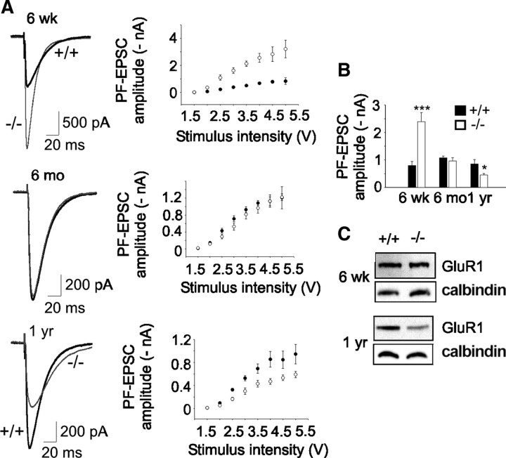
Altered PF-EPSCs in β-III−/− mice. A, Left, Representative EPSC waveforms (4 V stimulus) from 6-week-old, 6-month-old, and 1-year-old WT and β-III−/− littermates. Right, Mean PF-EPSC amplitudes versus stimulus intensity shows consistent differences between WT and β-III−/− cells at different stimuli. B, Mean peak amplitude of EPSCs at 4 V stimulus shows changes in PF-evoked currents with age in β-III−/− cells (6 weeks: N = 3, n = 10, p = 0.001; 6 months: WT, N = 2, n = 10; β-III−/−, N = 2, n = 9, p = 0.44; 1 year: WT, N = 1, n = 6; β-III−/−, N = 2, n = 11, p = 0.05). C, Western blot analysis shows loss of GluR1 in 1-year-old but no change in 6-week-old β-III−/− mice.

