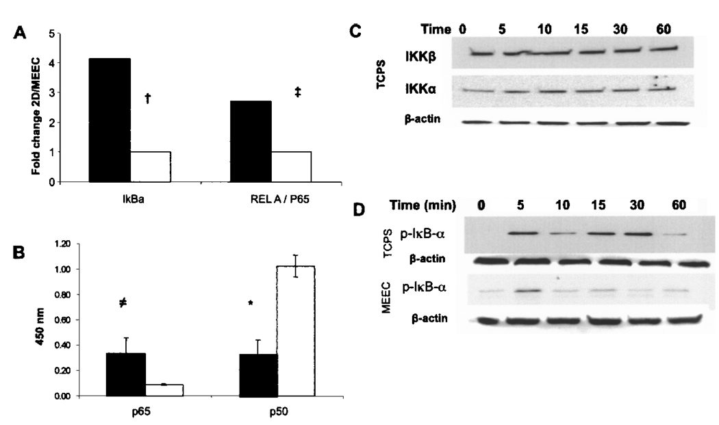
Figure 1.
Matrix embedding influences TNF-α-induced NF-κB activation in endothelial cells. (A) Real-time PCR fold change (ΔΔCt) values of IκBα, and REL-A (p65) for MEEC (opened histogram) and EC-TCPS (closed histogram) stimulated for 6 h with 5 ng/ml TNF-α. (B) Protein levels in the EC nucleus of p65 and p50, after 6-h stimulation with 5 ng/ml TNF-α in MEEC (opened histogram) and EC-TCPS (closed histogram). (C) Western blots of phosphorylated IKKα/β in EC-TCPS (levels were not detected in MEEC). (D) Western blots of phosphorylated IκBα over time in EC grown on TCPS. Western blots are representative for three independent experiments; equal loading was verified with β-actin.†p < 0.002, ‡p < 0.02, ≠p < 0.008, *p < 0.0001.

