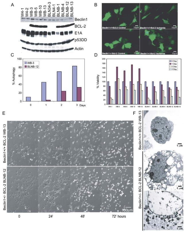
Figure 6. beclin1 haploinsufficiency impairs survival in ischemia in an apoptosis-defective background.
A: Western blot analysis of Beclin1, BCL-2, E1A, p53DD, and actin in beclin1+/+ and beclin1+/− iBMK cell lines with and without BCL-2 expression.
B: beclin1 haploinsufficiency impairs autophagy. beclin1+/+ and beclin1+/− iBMK cells expressing BCL-2 (WB-3 and BLNB-12) were transfected with the EGFP-LC3 expression vector and at 24 hr posttransfection were subjected to 0 and 24 hr of ischemia and were monitored for membrane translocation by fluorescence microscopy. Representative fields are shown.
C: Quantitation of EGFP-LC3 translocation (% autophagy) in WB-3 and BLNB-12 in B following 0, 1, 2, and 3 days in ischemia.
D: beclin1 haploinsufficiency impairs viability in ischemia only when apoptosis is inhibited. beclin1+/+ and beclin1+/− iBMKs with and without BCL-2 were incubated in ischemic conditions, and viability was assessed by trypan blue staining at the indicated times.
E: Time course of morphological alterations due to beclin1 haploinsufficiency in iBMK cells expressing BCL-2 (WB-13 and BLNB-12). beclin1+/+ cells expressing BCL-2 proliferate, followed by cellular condensation, whereas beclin1+/− cells expressing BCL-2 display diminished capacity to sustain proliferation and show accelerated necrosis.
F: EM (5000×) of beclin1+/+ BCL-2 (WB-13) and beclin1+/− BCL-2 (BLNB-12) following 7 days in ischemia. Note the condensed morphology of WB-13 and the vacuolated and necrotic morphology of BLNB-12.

