
Figure 6.
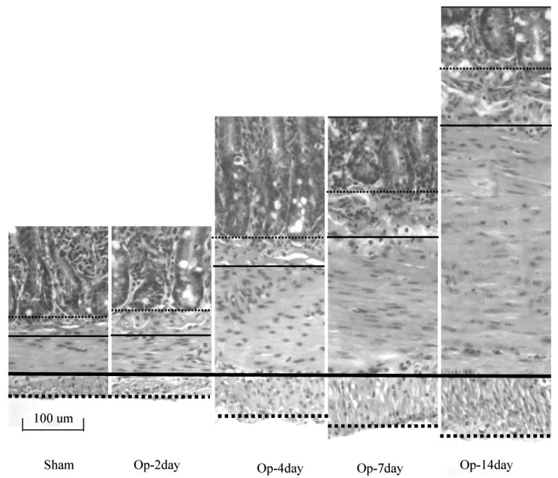
The typical pattern of the muscle layer proliferation and submucosa thickning. The thick dense-line, the thin dense-line, the thick dot-line and thin dot-line indicated the interface of two muscle layers, outer bordering of longitudinal muscle layer, the interface between muscle layer and submocosa layer, and the interface between submucosa layer and mucosa layer. The submucosa and muscle layers increased the thickness after the obstruction, the circumferential muscle layer increased much more than the longitudinal muscle layer.