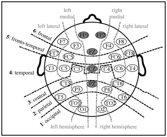
Figure 6.
Schematic representation of the electrode montage and the factors used in analyses. Six levels of the anterior/posterior factor, two levels of the lateral/medial factor, and two levels of the hemisphere factor are indicated. Electrodes in gray were not used in the primary analyses reported in the text.

