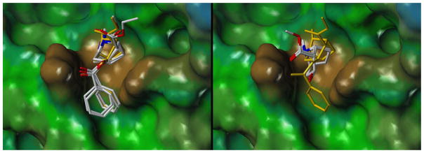
Figure 3.
Binding poses obtained for the “focused set” of cocaine metabolites (top view, cocaine is displayed in yellow). For clarity, the left panel contains the larger metabolites whereas the right panel shows the smaller compounds ecgonine and ecgoninemethylester alone. The surface of the antibody model is colored according to hydrophobicity (brown: more hydrophobic; green: less hydrophobic).

