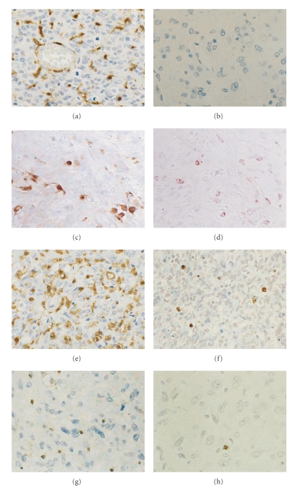
Figure 4.
Thymidine phosphorylase immunohistochemistry in human gliomas. Glioblastoma shows intense immunoreaction for thymidine phosphorylase both in tumor and endothelial cells (a). Diffuse astrocytoma shows no expression (b). Some of the tymidine phosphorylase positive cells (c) are macrophages ((d) serial section of (c)). Thymidine phosphorylase positive glioblastoma (e) reveals a high apoptotic index ((f) serial section of (e)), while Thymidine phosphorylase negative glioblastoma (g) reveals a low apoptotic index ((h) serial section of (g)). Original magnification ×200.

