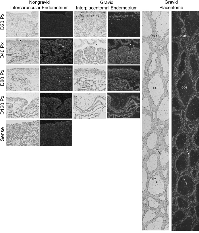
FIG. 5.
In situ hybridization analysis of S1PR1 mRNA in sections of nongravid and gravid uteri collected from unilaterally pregnant ewes on Days 20, 40, 80, and 120 of gestation. A composite image of a Day 80 placentome is shown (right panel). Corresponding bright field and dark field images of representative endometrial cross sections are shown. A section from Day 80 nongravid intercaruncular endometrium hybridized with radiolabeled sense cRNA probe served as a negative control. LE, endometrial luminal epithelium; GE, endometrial glandular epithelium; scST, endometrial stratum compactum stroma; Tr, placental trophectoderm; PL, placental chorion; CAR, caruncular tissue; COT, cotyledonary tissue; BV, blood vessel. Representative images are shown from independent samples (n = 4). Width of each field is 940 μm.

