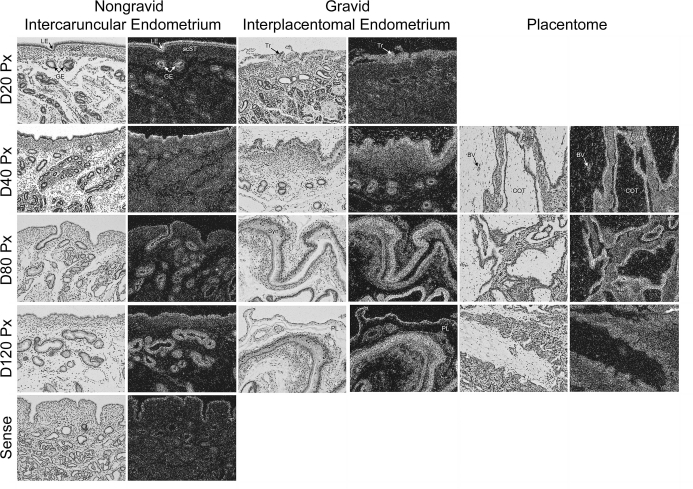
FIG. 9.
In situ hybridization analysis of ADAMTS1 mRNA in sections of nongravid and gravid uteri collected from unilaterally pregnant ewes on Days 20, 40, 80, and 120 of gestation. Corresponding bright field and dark field images of representative endometrial cross sections are shown. A section from Day 80 nongravid intercaruncular endometrium hybridized with radiolabeled sense cRNA probe served as a negative control. LE, endometrial luminal epithelium; GE, endometrial glandular epithelium; scST endometrial stratum compactum stroma; Tr, placental trophectoderm; BV, blood vessel; CAR, caruncular tissue; COT, cotyledonary tissue; PL, placental chorion; Syn, epithelial syncytia of the placentome. Representative images are shown from independent samples (n = 4). Width of each field is 940 μm.

